 | ||||

Mannitol Salts Agar (MSA) Specialized Media - P2
Page last updated: 4/2016
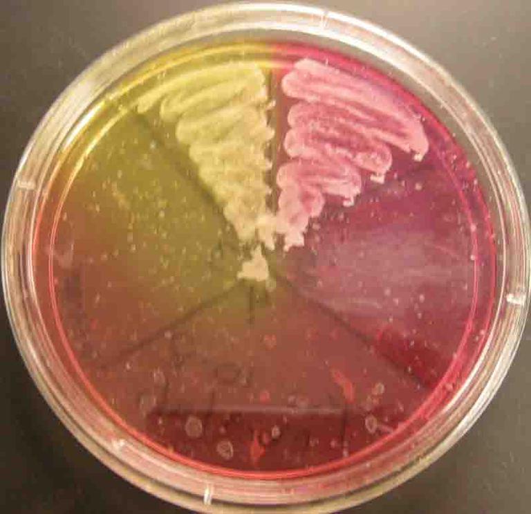
Plat of Mannitol Salt agar. On right side, plate is growing
Staphylococcus aureus, a pathogenic mannitol fermenter (yellow). On left, plate is growing non-mannitol fermenter
S. epidermidis.
Click here for more images of Mannitol Salt Agar.
You have free access to a large collection of materials used in a college-level introductory microbiology course. The Virtual Microbiology Classroom provides a wide range of free educational resources including PowerPoint Lectures, Study Guides, Review Questions and Practice Test Questions.

Button")
SCIENCE PHOTOS
SPO VIRTUAL CLASSROOMS
Mannitol Salt Is Differential
This growing medium is also differential because it contains a dye that identifies types of Staphylococcus that produce an organic acids from mannitol fermentation
(eating mannitol, a type of alcohol).
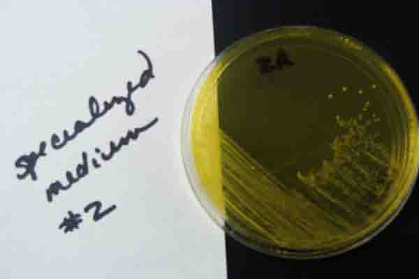
The bacterial waste products generated, organic acid metabolites, change the pH indicator in MSA from red to bright yellow. Pathogenic Staph, such as Staphylococcus aureus, are mannitol fermenters, and when growing on Mannitol Salt Agar, their wastes turn the MSA a bright yellow color.
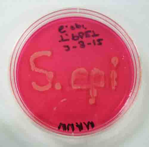
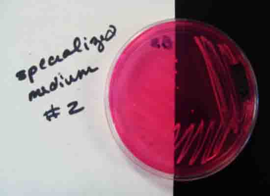
PHOTOS OF MANNITOL SALT AGAR (MSA): 1. Sterile plate of Mannitol Salt agar; 2. "Staph epi: written on MSA with the bacteria Staphylococcus epidermidis, a halophilic, non-mannitol fermenter; 3. A control plate of Mannitol Salt. Several different bacteria were plated, but only the halophiles grew. Staph aureus (top left, medium turned yellow) and Staph epi (top right, medium stayed pink); 4. Staph aureus plated on MSA; 5.Staph epi plated on MSA. Click on strip to enlarge photos.
Click here for more photos.

Importance of Identifying S. epi versus S. aureus
Staphylococcus epidermidis
S. epi bacteria is normally found on our skin and mucus membranes. Like all normal flora, it takes up space and resources, making difficult for other non-normal flora bacteria to grow. Because of their slime layer, and ability to form biofilms, S. epi can be problematic, as they are prone to build up on internal plastic medical devices.
Bacterial Virulence
Think of each little bacterium as having a tiny backpack that can be used to hold weapons called virulence factors. Virulence factors are features of a microbe that help it cause disease...weapons. Bacteria that do not often cause disease, such as normal flora, have fewer weapons in their backpack than do pathogens. The more pathogenic, typically the more weapons.
Staphylococcus aureus
S. aureus, the pathogen that has given rise to MERSA (Methicillin-resistant-Staphylococcus-aureus), has additional virulence factors, beyond just a slime layer. S. aureus
produces a number of enzymes such as coagulase that clots plasma and protects the bacterium. Another enzyme, hyaluronidase, helps the bacteria spread. And the enzyme beta-lactamase enables some S. aureus to resist some antibiotics. In addition to making a variety of enzyme virulence factors, strains of S. aureus also produces a number of toxins. A whole arsenal of weapons!
PAGE 2 < Back to Page 1


By contrast, nonpathogenic Staph such as Staphylococcus epidermidis (a.k.a. Staph epi), the normal flora that grows on human skin, does not ferment mannitol. When Staph epi grows on Mannitol Salt, the naturally orange-pink color of the agar doesn’t change, since S. epidermidis doesn’t eat mannitol or produce the resulting organic acid wastes.
VIDEO How to Interpret Mannitol Salt Agar
| ||||||
SPO is a FREE science education website. Donations are key in helping us provide this resource with fewer ads.
Please help!
(This donation link uses PayPal on a secure connection.)